Jan 27 2016
Bioengineering researcher Keith Markolf frequently gets this question when colleagues visiting from around the world first lay eyes on the 8-foot-tall, pumpkin-orange behemoth dominating the UCLA Orthopaedic Biomechanics Laboratory at UCLA’s Rehabilitation Center.
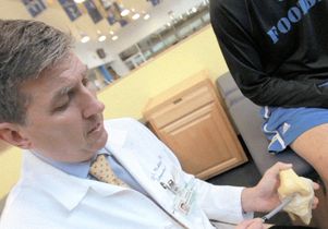
 Dr. David McAllister, with a member of the UCLA football team whose knee injury he treated, pointing to the ACL in a knee model.
The industrial robot, diverted from a thankless job in an auto assembly plant in Detroit, has taken on a second life as an explorer into the workings of the human knee.
The hulking robot thumps away incessantly, applying hundreds of pounds of force to a cadaver knee specimen implanted with a custom-designed sensor that measures forces in a knee ligament — a technique used nowhere else in the world but in this laboratory. Pairing this setup with a computer program that analyzes every move, Markolf and his collaborators at UCLA’s David Geffen School of Medicine — bioengineer Daniel Boguszewski and orthopaedic surgeon Dr. David McAllister — are shedding new light on how the knee works, how it gets injured and how best to repair it.
“We need a big robot to simulate big forces,” Markolf explained.
It is estimated that our knees absorb two to three times our body weight while we’re walking, and almost twice that during intense sports activities. This means that a leisurely walk or climbing up or down stairs can put up to 360 pounds of force on the knees of a person who weighs just 120 pounds. Add speed and impact during sports and the numbers multiply: A 215-pound football player’s knees can get clobbered with forces approaching 1,300 pounds when running at full speed.
Standing up to these forces on our knees is an undergirding of four small but strong bands of ligament tissue that strap together the large bones of our upper and lower legs. These ligaments enable us to stand up and move around with stability and grace instead of wobbling and collapsing like loose-jointed marionettes.
In recent years, the UCLA researchers have been focusing much of their work on the anterior cruciate ligament (ACL), the most important and most vulnerable of the knee ligaments. ACL injuries — sprains and the more serious ruptures, in which the ligament is torn — number approximately 200,000 every year in the United States, and they’re skyrocketing. Most of those injured are playing sports, from young soccer players to professional athletes. Females are six to 12 times more likely than males to tear their ACL, for reasons that remain a mystery.
“The ACL is a very complex ligament in terms of its geometry,” said Markolf. “It has different bands within its structure, and some of the bands become tight in full extension, and some become tight when the knee flexes. Overall, the ACL is extremely important in terms of stabilizing the knee. When the ligament is ruptured, the knee moves in ways that it didn’t before — in ways it shouldn’t move.”
Until recently, most research on knee injuries has been based on watching game films, said Boguszewski, who did his share of scrutinizing footage frame-by-frame before teaming up with a robot.
“You’d be trying to identify how the injury occurred based on a film of a football game,” he said. “But you wouldn’t really know how or exactly when the ligament ruptured. All you could see was how and when the player reacted.”
With their robotic testing system, the UCLA researchers are able to simulate potential injury scenarios and directly measure forces in the ACL that can lead to rupture. One common culprit is changing direction while running: When a football or soccer player running down the field plants his foot hard to change direction, his knee can buckle as his upper leg moves in one direction while his lower leg moves in another. Also tough on the knees are the twists and jumps of basketball players and the hard landings gymnasts make in high-velocity dismounts from a bar.
Studying ACL injuries with a robot at UCLA
McAllister, as lead physician at the UCLA Sports Medicine Center, is a frontline responder to injuries suffered by any of UCLA’s more than 700 student-athletes. He has treated dozens of Bruins knocked out of games with ACL sprains and ruptures. Injuries like these could end a sports career, but advances in surgical reconstruction and rehabilitation are changing this. Every year, McAllister performs hundreds of knee reconstruction surgeries — in which a section of tendon attached to the kneecap or a portion the lower leg’s hamstring tendon is affixed to the bones to craft a replacement ligament — for athletes and non-athletes alike.
But reconstruction isn’t always a permanent fix. Sometimes the replacement ligament fails outright when athletes return to high-intensity sports activities. And even when surgery succeeds, ACL reconstruction can be accompanied by future osteoarthritis and cartilage damage. An important goal of the UCLA lab’s biomechanical studies is to develop treatment strategies that will best stabilize the knee to prevent further degeneration in future years.
McAllister and his collaborators are applying their robot-powered research to scientifically determine which approaches to reconstruction used by surgeons around the world are actually working and which ones aren’t.
“A few years ago, a new surgical technique using double ACL graft bundles was proposed as possibly being better than what surgeons were doing at that time,” McAllister said. “We tested it and found it wasn’t better and, in fact, has some distinct disadvantages. This newer technique has now fallen out of favor in North America.”
The UCLA lab’s studies also provide important insights into the mechanisms that lead to injuries, as well as how to best rehabilitate the knee after ACL reconstruction surgery.
“What we learn has clinical relevance,” McAllister said. “I can it use immediately with patients — and with confidence that it will work."