Treatment choices for individuals with acute myeloid leukemia (AML), an extremely aggressive type of disease, are influenced by several specific genetic characteristics of the disease; however, at the time of diagnosis, this information is unavailable. On the other hand, early detection of these genetic abnormalities is essential for delivering patients with tailored treatment.
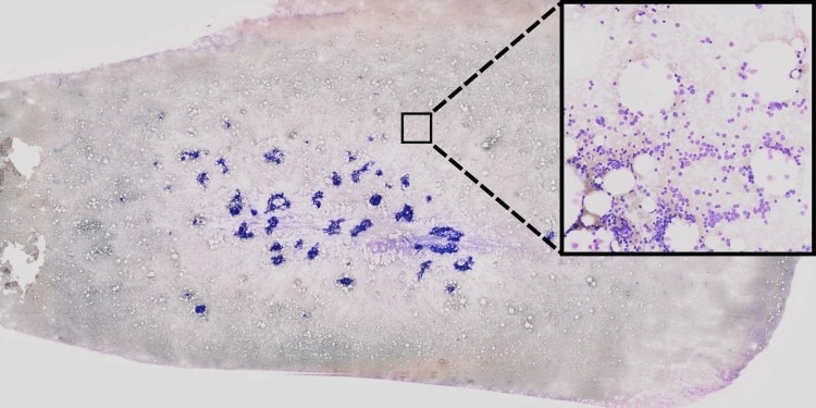
 AI-based processing of bone-marrow smears to support leukemia diagnosis. Using so-called unsupervised learning methods, single-cell images (center) are extracted from extremely high-resolution image data (left). Then, using neural networks, these cell images are examined to check for any visual anomalies which might have a genetic origin. Areas which are important for the decision of the neural network are highlighted in color by using so-called “explainable AI” strategies (right). Image Credit: AG Risse
To predict such abnormalities, there is a huge need for affordable, quick, and widely available assays, as genetic testing is costly and time-consuming.
In a recent study, a group of doctors and IT experts from the University of Münster and the University Hospital Münster demonstrated how different genetic features can be predicted using artificial intelligence (AI) based on high-resolution microscopic images of bone marrow smears.
Consequently, future treatment decisions can be taken promptly on the day of diagnosis, without waiting for genetic tests. This allows for more precise treatment planning. The findings have been released in the Blood Advances journal.
Using this innovative technique, complete bone marrow smears from over 400 AML patients were scanned at extraordinarily high resolution (multi-gigabyte), directly yielding the genetic abnormalities. The average resolution of the scans was 270,000 by 135,000 pixels, and each image weighed several terabytes. More than two million single-cell images might be extracted from this massive collection.
We developed a new type of Deep Learning method, fully automatic, which was trained for a complex task by means of machine learning technology. In our case, the basic algorithm can automatically recognize the genetic features and the very fine patterns in big cytological images. The method then filtered the single-cell images into categories of different cell types, and it also showed any genetic aberrations.
Benjamin Risse, Professor, University of Münster
Risse added, “Interestingly, several patterns recognized by the algorithm cannot be identified by human observers. This is for example because the patterns may be too faint or because extremely fine textures are involved which remain hidden to the human eye, despite excellent imaging.”
The method’s end-to-end AI pipeline, which allows for monitoring of the (interim) outcomes and minimizes the human preliminary work sometimes required for machine learning, is one of its main advantages.
A mix of self-supervised, supervised, and so-called unsupervised learning processes enables this. Instead of requiring any manual data selection at all, the first two steps attempt to automatically extract pertinent material from the image data.
Using a so-called incremental approach, we carried out intermediary steps with a human expert to examine the images. This is necessary for example in cell images categorized as problematic.
Dr. Linus Angenendt, Head, Personalized Cancer Therapy and Digital Medicine working group, Münster University Hospital
For example, improper staining might result in problematic cell images. The trained model was then assessed as a test cohort using an independent dataset that included approximately 440,000 single-cell images from an additional 70 patients.
The novel approach aids in the early stages of a leukemia patient’s diagnostic clarification process by identifying potential genetic aberrations that could represent the cause of the disease, even though it cannot completely replace genetic studies. When there is no time to wait for the whole genetic test, this would be especially useful in the case of aggressive diseases.
When it comes to providing patients with malignant diseases with individualized treatment suggestions, the researchers are optimistic that digital techniques and artificial intelligence will play a bigger role in the future for vast medical databases. The study provides a solid foundation for it, for instance in the creation of comparable methods for treating other bone marrow diseases.
Funding for the study was provided by the German Research Foundation and the European Union.
Journal Reference:
Kockwelp, J., et. al. (2023) Deep learning predicts therapy-relevant genetics in acute myeloid leukemia from Pappenheim-stained bone marrow smears. Blood Advances. doi:10.1182/bloodadvances.2023011076