May 11 2021
The World Health Organization reports that, in recent times, breast cancer has exceeded lung cancer to become the most common type of cancer worldwide.
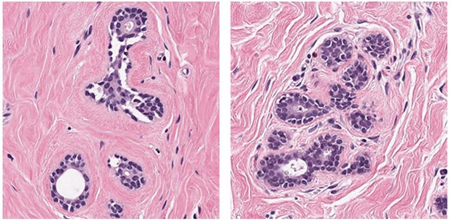
 AI algorithms performed well on easier breast cancer image patches, like these. Image Credit: Petrick et al., doi 10.1117/1.JMI.8.3.034501
The BreastPathQ Challenge was started at SPIE Medical Imaging 2019 to progress the fight against breast cancer and to help develop a computer-aided diagnosis for evaluating breast cancer pathology.
The participants of BreastPathQ Challenge were given a task to design an automated technique for examining microscopy images of breast tissue and rating them as per their tumor cell content, to offer a trustworthy assessment score.
As stated in SPIE’s Journal of Medical Imaging (JMI), the task produced promising outcomes that denote a path toward combining artificial intelligence (AI) to simplify clinical evaluation of breast cancer.
Medical Imaging for Neoadjuvant Treatment
Treatment for aggressive or huge breast cancers has usually resorted to mastectomy as the most reliable treatment. But a therapy called “neoadjuvant treatment” can lead to decreased tumor density, size, and spread, thereby making patients as candidates for breast-conserving surgery instead of mastectomy.
Medical imaging enables doctors to evaluate the impacts of neoadjuvant treatment. Generally, while the processes of examining medical images for cancer detection are performed manually and depend on expert interpretation of complicated tissue structures, machine-learning algorithms for determining cancer may enhance the efficiency and reliability of such processes.
Besides decreasing variability, which is innate to human pathologists, completely automated techniques like these are anticipated to raise the speed of image analysis.
Intensive Focus, International Effort
From around the world, 39 research groups from 12 different countries were involved in the BreastPathQ Challenge. In total, 100 algorithms were designed, validated, and tested. The teams could make a comparison of their algorithms with those of others from government, industry, and academia, and as structured by the Grand Challenge framework, which needs a shared set of source data.
An ensemble of machine-learning algorithms was used by the majority of the groups rather than restricting themselves to an individual AI architecture. Top algorithms exhibited performance at levels equivalent to the pathologists who offered the reference standards for the study, and the best functioning algorithm slightly exceeded the scores of the pathologists.
Normally, the algorithms worked well on simpler patches of images but were difficult to work on the hard patches—those for which AI would be particularly advantageous to pathologists.
The BreastPathQ Challenge was a successful one since the organizing committee assembled experts from various fields.
Nicholas Petrick, deputy director for the Division of Imaging, Diagnostics and Software Reliability in the US FDA Center for Devices and Radiological Health, and representative for the BreastPathQ Challenge Group, stated that advance collaborative groundwork implied that participants could move rapidly and efficiently to deal with the task, access the data set, and develop their algorithms.
Journal Reference:
Petrick, N., et al. (2021) SPIE-AAPM-NCI BreastPathQ challenge: an image analysis challenge for quantitative tumor cellularity assessment in breast cancer histology images following neoadjuvant treatment. Journal of Medical Imaging. doi.org/10.1117/1.JMI.8.3.034501.