Jul 28 2020
A new study by researchers from UPMC and the University of Pittsburgh has reported the highest-ever accuracy so far in the identification and characterization of prostate cancer with the help of an artificial intelligence (AI) program.
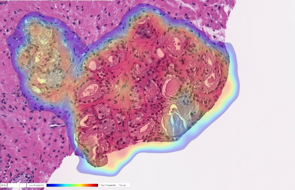
 Prostate biopsy with cancer probability (blue is low, red is high). This case was originally diagnosed as benign but changed to cancer upon further review. The AI accurately detected cancer in this tricky case. Image Credit: Ibex Medical Analytics.
The study was recently published in The Lancet Digital Health.
Humans are good at recognizing anomalies, but they have their own biases or past experience. Machines are detached from the whole story. There’s definitely an element of standardizing care.
Rajiv Dhir, MD, MBA, Chief Pathologist and Vice Chair of Pathology, UPMC Shadyside Hospital
Dhir is the senior author of the study and also a professor of biomedical informatics at the University of Pittsburgh.
Dhir and his team trained the AI to identify prostate cancer by providing images from over a million parts of stained tissue slides obtained from patient biopsies. Expert pathologists labeled each image to train the AI to differentiate between abnormal and healthy tissue.
The algorithm was then tested on another set of 1,600 slides obtained from 100 consecutive patients observed at UPMC for suspected prostate cancer.
As part of the tests, the AI exhibited 98% sensitivity and 97% specificity in the detection of prostate cancer, which is considerably higher compared to figures reported earlier for algorithms that work using tissue slides.
Moreover, this algorithm is the first one to have applications beyond cancer detection, achieving high performance for tumor grading, sizing, and invasion of the adjacent nerves. All these features are clinically important and essential as part of the pathology report.
In addition, the AI flagged six slides not noted by the expert pathologists.
However, Dhir noted that this does not mean that the machine is better than humans. For instance, while assessing these cases, the pathologist could just have observed sufficient evidence of malignancy in other areas of that patient’s samples to recommend treatment. But in the case of less experienced pathologists, the algorithm could serve as a secure way to arrest cases that could be missed otherwise.
Algorithms like this are especially useful in lesions that are atypical. A nonspecialized person may not be able to make the correct assessment. That’s a major advantage of this kind of system.
Rajiv Dhir, MD, MBA, Chief Pathologist and Vice Chair of Pathology, UPMC Shadyside Hospital
Although the study outcomes look promising, Dhir warns that new algorithms must be trained to recognize cancer of different types. The pathology markers are not universally applicable to all tissue types. However, it was not clear why that could not be performed to adapt this technology for breast cancer, for instance.
Liron Pantanowitz, MBBCh, from the University of Michigan; Gabriela Quiroga-Garza, MD, from UPMC; Lilach Bien, Ronen Heled, Daphna Laifenfeld, PhD, Chaim Linhart, Judith Sandbank, MD, Manuela Vecsler, from Ibex Medical Analytics; Anat Albrecht-Shach, MD, from Shamir Medical Center; Varda Shalev, MD, MPA, from Maccabbi Healthcare Services; and Pamela Michelow, MS, and Scott Hazelhurst, PhD, from the University of the Witwatersrand are the additional authors of the study.
This study was financially supported by Ibex, which also developed this commercially available algorithm.
Journal Reference:
Pantanowitz, L., et al. (2020) An artificial intelligence algorithm for prostate cancer diagnosis in whole slide images of core needle biopsies: a blinded clinical validation and deployment study. The Lancet Digital Health. doi.org/10.1016/S2589-7500(20)30159-X.