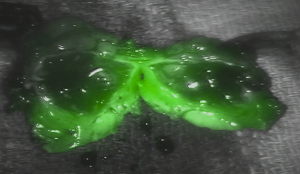
Surgeons at the USMD Hospital have used florescence imaging techniques for the first time while performing robotic kidney surgery. Using this technique, provides superior view of the anatomy being operated.
 flourescence image of body tissues
By using fluorescence imaging, the surgeon is able to see the anatomy better with the naked eye and perform precise operation of the tumour, leaving the surrounding healthy tissues untouched. From the view point of the patient, robotic surgery results in lesser pain, faster recovery times and lesser post-operative complications.
One of the surgeons at the hospital pointed out that USMB was the first to offer fluorescence-aided robotic surgical techniques and that the hospital has completed more than 4500 robotic procedures. For the fluorescence technique they require a specially designed camera and endoscopes that capture tissue and neighbouring blood vessel images. The fluorescence dye is injected into the body in order to highlight the tissue when activated by light from near-infrared region. For better clarity real-time images of the surgical field are provided to the surgeon, he can switch from these images to the fluorescence images as and when required. Physicians are in a position to distinguish between malignant tissue and benign tissue more accurately through this technique.
The technique when used along with the da Vinci surgical system will be extended on surgeries other than that of the kidney at the USMD, following the clearance received from FDA by Intuitive, the maker of da Vinci Surgical System. The da Vinci Surgical Robot makes use of the minimally invasive surgical technique that results in faster recovery while performing procedures such as Bariatric cases, gynecologic surgeries and prostatectomies. The da Vinci system is equipped with high-definition cameras that provide three dimensional views to the surgeon that enable superior control of the surgical instruments during surgery.