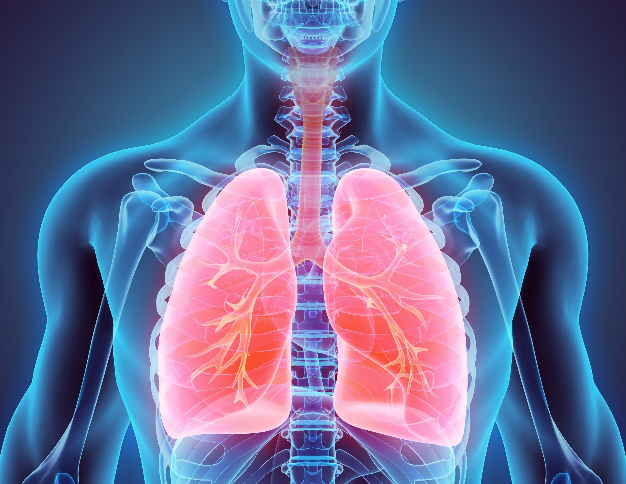
A team of researchers from Imperial College London, in collaboration with Imperial College Healthcare NHS Trust and international partners, has harnessed the power of artificial intelligence (AI) to analyze medical scans for lung cancer assessment.

Image Credit: MDGRPHCS/Shutterstock.com
This innovative approach, termed a 'virtual biopsy,' offers a non-invasive alternative to traditional biopsy methods, providing critical insights into the chemical composition of lung tumors directly from imaging data.
The significance of this development cannot be overstated for industries reliant on cutting-edge medical technologies, including healthcare providers, medical imaging equipment manufacturers, and pharmaceutical companies.
By enabling precise classification of lung cancer types and predicting disease progression without the need for invasive tissue sampling, this technique promises to streamline diagnostic workflows, reduce healthcare costs, and accelerate the personalization of treatment strategies.
Central to this achievement is the integration of AI with computed tomography (CT) scans to create a sophisticated analysis model that the researchers have named Tissue-Metabolomic-Radiomic-CT (TMR-CT).
This model leverages deep learning algorithms to identify correlations between the visual features of CT scans and the metabolic profiles of tumors, traditionally obtained through metabolomic profiling of biopsy samples.
We’ve developed a system that merges CT scans with the chemical makeup of tumours and normal lung tissue. This allows us to classify lung cancer types and, importantly, provides reliable predictions about patient outcomes.
Marc Boubnovski Martell, First-author and Imperial PhD Candidate
The approach was rigorously validated using data from a cohort of 48 lung cancer patients who underwent both CT scanning and detailed metabolomic analysis of their tumors at the University Hospital Reina Sofia in Córdoba, Spain.
The TMR-CT model's efficacy was further demonstrated in a larger sample of 723 lung cancer patients treated across several prominent UK hospitals. These patients had undergone CT scans but lacked corresponding metabolomic data.
Remarkably, the TMR-CT model not only classified lung cancer types with high accuracy but also provided reliable prognostic predictions, outperforming conventional CT-based diagnostic methods.
This innovation opens new horizons for the early detection and treatment of lung cancer, which remains a leading cause of cancer-related mortality worldwide. By eliminating the need for physical biopsy samples, this method stands to benefit patients for whom traditional biopsy procedures are not viable, thereby ensuring timely and appropriate treatment interventions.
Looking ahead, the research team, led by Professor Eric Aboagye, envisions expanding the application of the TMR-CT model to other cancers, such as brain, ovarian, and endometrial cancers, where obtaining biopsy samples poses similar challenges.
The ultimate goal is to integrate this AI-driven diagnostic tool into commercial medical imaging scanners, transforming the landscape of cancer diagnostics and treatment planning.
This research shows the potential of using CT scans to gain a deeper, more nuanced understanding of tissue and tumour chemical composition, that has until now only been accessible through direct tissue sampling. This method could prove particularly beneficial in countries like the UK, where lung cancer prevalence is high, and potentially transform diagnostic and treatment protocols.
Professor Eric Aboagye, Department of Surgery and Cancer, Imperial College London
This study, published in the journal npj Precision Oncology, successfully highlights the potential role of AI in enhancing a practitioner’s – radiologist’s, respiratory physician’s, or oncologist’s – ability to determine histology subtype classification, as well as prognosis, using algorithms derived from current work.
It not only marks a significant step forward in the field of precision oncology but also underscores the critical role of interdisciplinary collaboration in harnessing the full potential of AI to address complex medical challenges.
References and Further Reading
-
Czyzewski, A. (2024) ‘virtual biopsy’ uses AI to help doctors assess lung cancer: Imperial News: Imperial College London, Imperial News. Available at: https://www.imperial.ac.uk/news/251593/virtual-biopsy-uses-ai-help-doctors/ (Accessed: 26 February 2024).
-
Boubnovski Martell, M. et al. (2024) ‘Deep representation learning of tissue metabolome and computed tomography annotates NSCLC classification and Prognosis’, npj Precision Oncology, 8(1). Available at: https://www.nature.com/articles/s41698-024-00502-3#Sec1. (Accessed: 26 February 2024).