Researchers from the University of Jyväskylä, in cooperation with the Institute of Biomedicine at the University of Turku and Nova Central Finland, have created an artificial intelligence tool designed for the automatic analysis of colorectal cancer tissue.
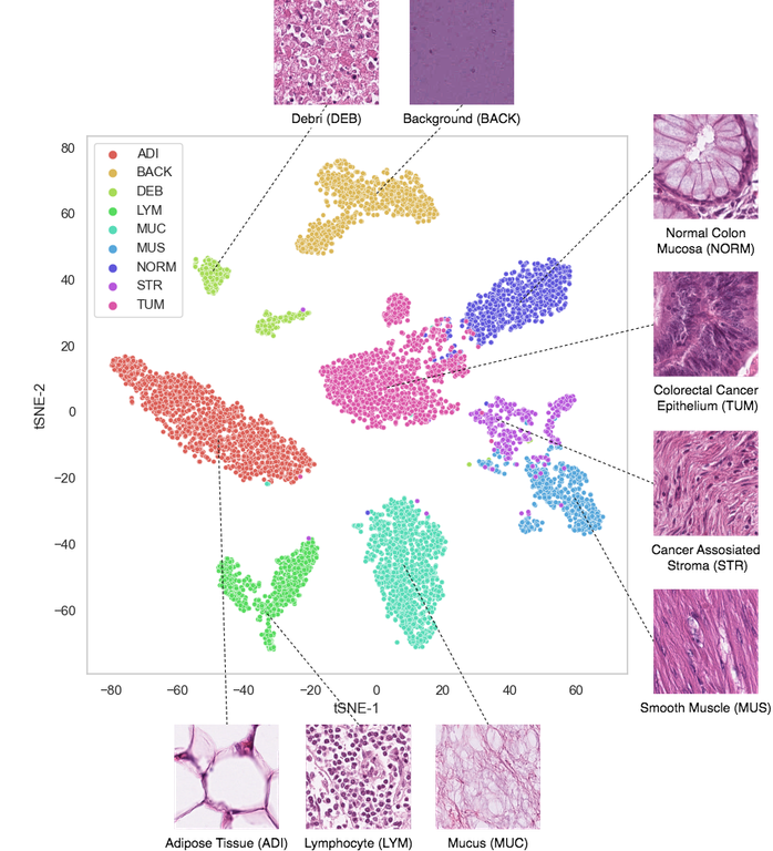
 Each dot represents an image piece from a cancer microscope slide. The AI system has automatically arranged them by similarity. Image Credit: University of Jyväskylä.
The improved neural network outperformed all prior solutions in this particular task. This neural network provides a quicker and more precise means of classifying colorectal cancer tissue images from microscope slides. As a result, it has the potential to substantially reduce the workload of histopathologists, ultimately leading to faster insights, prognoses, and diagnoses in the field of colorectal cancer.
Public Accessibility for Future Advancements
In a notable commitment to open science and collaborative progress, the researchers are offering public access to the trained neural network tool. This enables further advancements by inviting scientists, researchers, and developers globally to refine their capabilities and investigate new applications.
By granting universal access, the aim is to fast-track breakthroughs in colorectal cancer research.
Fabi Prezja, Doctoral Student, University of Jyväskylä
Stringent Clinical Validation is Imperative
It is vital to adopt AI in clinical settings sensibly, though the results are assuring. The caliber and diversity of medical data play a major role in the achievement of AI-driven methods. The AI solutions undergo severe clinical validation, though similar to routine clinical implementation, ensuring their outcomes consistently align with clinical standards.
Journal Reference:
Prezja, F., et al. (2023). Improved accuracy in colorectal cancer tissue decomposition through refinement of established deep learning solutions. Scientific Reports. doi.org/10.1038/s41598-023-42357-x