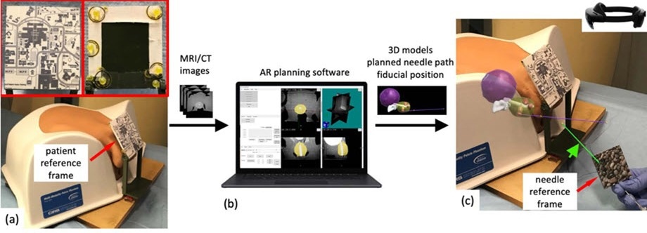
A HoloLens AR system has been demonstrated by scientists that allows precise and adaptable needle guidance for transperineal prostate interventions like focal laser ablation, biopsy, and brachytherapy.

The HoloLens AR system superimposes anatomical features generated from MRI or CT scans onto the patient for real-time needle guidance in prostate interventions. Image Credit: Li et al., doi.org/10.1117/1.JMI.10.2.025001
As per the US Center for Disease Control and Prevention, among men, prostate cancer is known to be the second chief cause of cancer death. One of the standard methods for the diagnosis and treatment of prostate cancer includes transperineal (TP) biopsy.
This includes the insertion of a needle via the perineum wall to gather tissue samples. New techniques available at present for TP biopsy generally consist of transrectal ultrasound and pre-operation MRI scan.
These images are fused and displayed on a monitor for the urologist, who then inserts the needle. The needle insertion could be executed free-hand or through a grid-based technique. However, since this method involves envisioning a 3D region in 2D, visualization and guidance of the needle can be difficult.
For this problem to be addressed, a research group headed by Dr. Ming Li from the US National Institutes of Health has suggested a method based on an augmented reality (AR) system known as HoloLens.
In their recent study reported in the Journal of Medical Imaging, the scientists report the precision and efficacy of their AR HoloLens system, which could be utilized to project the MRI and ultrasound image directly onto the patient, thereby helping direct a needle to its target.
The current methods have certain limitations to them. Robot-assisted guidance is costly and adds procedural time, while other methods require a certain path for the needle that leaves out the outer reaches of the prostate. These problems are solved using the HoloLens AR system, which provides the doctor with the ability to use a free-hand approach.
Dr. Ming Li, Staff Scientist, US National Institutes of Health
The HoloLens AR system makes use of a volumetric 3D scan, like an MRI scan, to make a view of the patient for the urologist. With the help of reference data from the patient, the MRI scan could be superimposed onto the patient properly.
This superimposed image is fed into the HoloLens goggles that have been worn by the urologist, who can see not only the patient but also the MRI of the patient aligned correctly. By moving their head, it is possible for them to view the image from different angles.
Also, HoloLens could be utilized to display the target tissue of the prostate, a preplanned needle path on the patient, and also the position of the needle in real time.
The image overlay accuracy and needle targeting accuracy were tested by the scientists for their AR system with the help of a 3D-printed phantom. Both planned-path and free-hand guidance techniques were utilized to guide needles into a gel phantom.
Subsequently, the placement errors for the needles were recorded by the scientists. They also utilized the system to offer soft-tissue markers onto the tumors of a human pelvis phantom.
They discovered that the placement errors linked to free-hand and planned-path guidance were alike. Furthermore, the soft tissue markers were all ingrained either into or close to the tumors.
The HoloLens has the potential to provide more flexibility than the current grid-based TP methods and can do so accurately. By providing a 3D immersive experience, the HoloLens AR system makes free-hand lesion targeting feasible. As needle procedures move from rectum to the perineum, AR systems could provide great clinical value to doctors and patients by solving the problems associated with prostate intervention procedures.
Dr. Ming Li, Staff Scientist, US National Institutes of Health
Journal Reference
Li, M., et al. (2023) HoloLens augmented reality system for transperineal free-hand prostate procedures. Journal of Medical Imaging. doi.org/10.1117/1.JMI.10.2.025001.