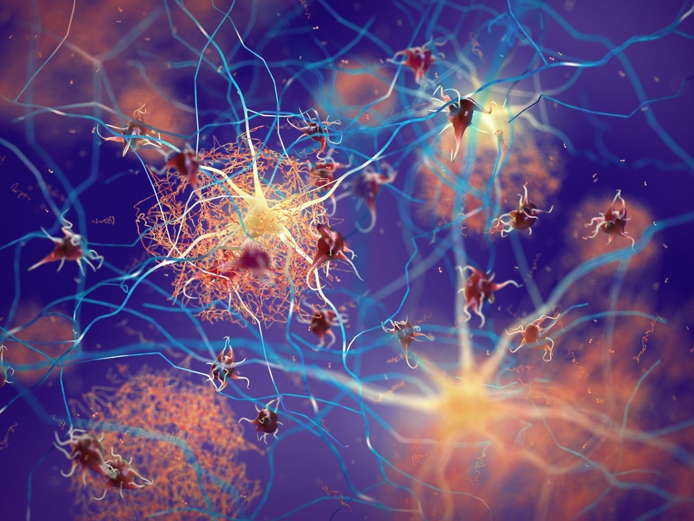
The human brain carries several clues regarding an individual’s long-term health—in fact, studies show that the brain age of a person is a more valuable and precise forecaster of health threats and future diseases when compared to their birthdate.

Image Credit: nobeastsofierce/Shutterstock.com
Currently, a new artificial intelligence (AI) model that examines magnetic resonance imaging (MRI) brain scans built by USC scientists can be employed to precisely capture cognitive decline in association with neurodegenerative diseases such as Alzheimer’s much earlier than other methods.
Aging of the brain is regarded as a dependable biomarker for the risk of neurodegenerative disease. This risk rises when an individual’s brain shows characteristics that seem “older” than anticipated for someone of that individual’s age. By tapping into the deep learning capacity of the new AI model of the team to examine the scans, the scientists can identify subtle brain anatomy markers, which are rather challenging to identify and associate with cognitive decline. The results—which were published on January 2nd, 2023 in the journal Proceedings of the National Academy of Sciences—provide an unparalleled insight into human cognition.
Our study harnesses the power of deep learning to identify areas of the brain that are aging in ways that reflect a cognitive decline that may lead to Alzheimer’s. People age at different rates, and so do tissue types in the body. We know this colloquially when we say, ‘So-and-so is forty, but looks thirty. The same idea applies to the brain. The brain of a forty-year-old may look as ‘young’ as the brain of a thirty-year-old, or it may look as ‘old’ as that of a sixty-year-old.
Andrei Irimia, Study Corresponding Author and Assistant Professor, Gerontology, Biomedical Engineering, Quantitative & Computational Biology and Neuroscience, USC Leonard Davis School of Gerontology
A More Accurate Alternative to Existing Methods
Irimia and his team gathered the brain MRIs of 4,681 cognitively normal participants. The participants include some of those who developed Alzheimer’s disease or mental decline in their future life.
With these data, they produced an AI model known as a neural network to forecast the ages of participants from their brain MRIs. Firstly, the scientists trained the network to generate elaborate anatomic brain maps that show subject-specific aging patterns. Then, they compared the observed brain ages—the biological ages—with the actual ages—which are the chronological ages—of study participants. The larger the difference between the two, the worse the cognitive scores of participants, reflecting the risk of Alzheimer’s.
The findings present that the model of the team can foresee the true (chronological) ages of mentally normal participants with 2.3 years average absolute error, which is around one year more precise than an already available, award-winning model for brain age estimation that employed a different neural network architecture.
“Interpretable AI can become a powerful tool for assessing the risk for Alzheimer’s and other neurocognitive diseases. The earlier we can identify people at high risk for Alzheimer’s disease, the earlier clinicians can intervene with treatment options, monitoring, and disease management.”
What makes AI especially powerful is its ability to pick up on subtle and complex features of aging that other methods cannot and that are key in identifying a person’s risk many years before they develop the condition.
Andrei Irimia, Study Corresponding Author and Assistant Professor, Gerontology, Biomedical Engineering, Quantitative & Computational Biology and Neuroscience, USC Leonard Davis School of Gerontology
Irimia also holds faculty roles with the USC Viterbi School of Engineering and USC Dornsife College of Letters, Arts, and Sciences.
Brains Age Differently According to Sex
The new model also shows sex-specific changes in how aging differs among brain areas. Some brain parts age quicker in males when compared to females, and vice versa.
Males have an increased risk of motor impairment owing to Parkinson’s disease and go through quicker aging in the brain’s motor cortex, a region accountable for motor activity. Results also reveal that typical aging might be comparatively slower in the brain’s right hemisphere among females.
An Emerging Field of Study Shows Promise for Personalized Medicine
The uses of this study extend much beyond disease risk assessment. Irimia pictures a world where the new deep learning methods developed as part of the study are employed to aid people in comprehending how quickly they are aging generally.
“One of the most important applications of our work is its potential to pave the way for tailored interventions that address the unique aging patterns of every individual,” Irimia added.
“Many people would be interested in knowing their true rate of aging. The information could give us hints about different lifestyle changes or interventions that a person could adopt to improve their overall health and well-being. Our methods could be used to design patient-centered treatment plans and personalized maps of brain aging that may be of interest to people with different health needs and goals,” he concludes.
Journal Reference:
Yin, C., et al. (2023) Anatomically interpretable deep learning of brain age captures domain-specific cognitive impairment. Proceedings of the National Academy of Sciences. doi.org/10.1073/pnas.2214634120.