Reviewed by Mila PereraSep 14 2022
As per a study published in Radiology, an AI device is very efficient at identifying pancreatic cancer on CT.

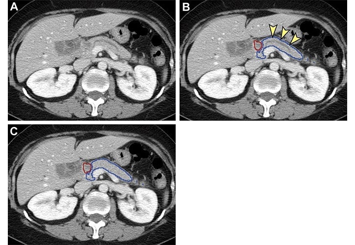
Analysis of nontumorous portion of pancreas with or without secondary signs of pancreatic cancer by classification models. Blue outline represents the portion of the pancreas analyzed with classification models. The tumor (red outline) was not identified by the segmentation model; thus, it was not analyzed by classification models. (A) Unannotated CT image in a patient with pancreatic head cancer. (B) Nontumorous portion of the pancreas shows secondary signs of pancreatic cancer (dilation of pancreatic duct with abrupt cutoff [arrowheads]) and was classified as cancerous by the classification models. (C) Nontumorous portion of the pancreas appeared normal and was classified as noncancerous after the dilated duct was replaced and imputed with surrounding normal-appearing pancreas parenchyma. Image Credit: Radiological Society of North America
Among cancers, pancreatic cancer has the lowest five-year survival rate. By 2030, it is predicted to become the second leading cause of cancer death in the US. As diagnosis deteriorates considerably after the tumor grows more than 2 cm, early prognosis is the ideal way to improve the dismal outlook.
Although CT is the primary imaging approach for pancreatic cancer diagnosis, it tends to miss around 40% of tumors under 2 cm. There is an immediate need for an efficient device to help radiologists improve pancreatic cancer detection.
Taiwanese scientists have investigated a computer-aided detection (CAD) tool that utilizes deep learning to identify pancreatic cancer. They demonstrated that the tool could differentiate pancreatic cancer from the noncancerous pancreas. The study, however, depended on the radiologists' manual identification of the pancreas on imaging.
The AI tool automatically found the pancreas in the new research. This indicated critical progress considering that the pancreas differs significantly in size and shape and borders several structures and organs.
Tool Has Potential to Detect Smaller Cancers Earlier
The scientists built the tool with an internal test set comprising 733 control participants and 546 patients with pancreatic cancer. The tool accomplished 96% specificity and 90% sensitivity in the internal test set.
The confirmation was pursued with a set of 1,473 individual CT exams carried out from institutes across Taiwan. In differentiating pancreatic cancer from controls, the tool attained 93% specificity and 90% sensitivity in that set. For identifying pancreatic cancers that are below 2 cm, the sensitivity was 75%.
The performance of the deep learning tool seemed on par with that of radiologists. Specifically, in this study, the sensitivity of the deep learning computer-aided detection tool for pancreatic cancer was comparable with that of radiologists in a tertiary referral center regardless of tumor size and stage.
Weichung Wang, Ph.D., Study Senior Author and Professor, National Taiwan University
Dr. Wang is also the director of the university’s MeDA Lab.
Dr. Wang stated that the CAD tool could offer much information to help clinicians, showing the area of suspicion to pace up radiologist interpretation.
The CAD tool may serve as a supplement for radiologists to enhance the detection of pancreatic cancer.
Wei-Chi Liao, MD., Ph.D., Study Co-Senior Author, National Taiwan University and National Taiwan University Hospital
The researchers have planned further studies. Specifically, they want to test the tool's performance in more varied populations. As the present study was retrospective, they would like to observe its functions in the future in practical clinical settings.
Journal Reference
Chen, P. T., et al (2022) Pancreatic Cancer Detection on CT Scans with Deep Learning: A Nationwide Population-based Study. Radiology. doi.org/10.1148/radiol.220152.