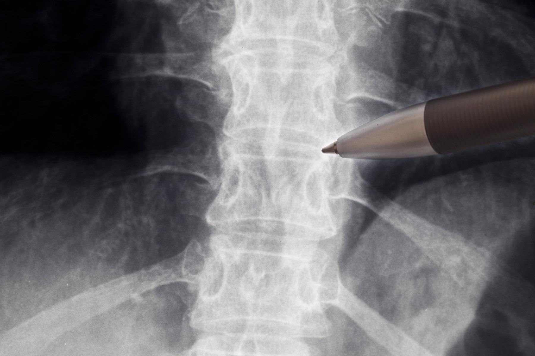
As medicine embraces machine learning, a new study shows that scientists could use artificial intelligence to forecast how cancer might influence the probability of spinal fractures.
 Using non-invasive tools like AI could help researchers better understand and treat bone fractures. Image Credit: Getty images.
Every year, more than 1.6 million cancer cases are identified in the United States, and around 10% of those people develop spinal metastasis, which occurs when the disease spreads from other parts of the body to the spine. One of the most serious clinical concerns that patients face is the possibility of spinal fractures as a result of these tumors, which can cause significant pain and spinal instability.
Spinal fracture increases the risk of patient death by about 15%. By predicting the outcome of these fractures, our research offers medical experts the opportunity to design better treatment strategies, and help patients make better-informed decisions.
Soheil Soghrati, Study Co-Author and Associate Professor, Department of Mechanical and Aerospace Engineering, The Ohio State University
While many of the changes the body experiences, when exposed to cancerous lesions, remain a mystery, scientists can gain a better understanding of what is going on in the spine via computer modeling, according to Soghrati.
The researchers detail how scientists trained an AI-assisted framework called ReconGAN to produce a digital twin, or a virtual reconstruction of a patient’s vertebra, in their article, which was published in the International Journal for Numerical Methods in Biomedical Engineering.
Unlike 3D printing, which includes turning a virtual model into a physical product, the idea of a digital twin entails generating a computer simulation of its real counterpart without actually physically making it. A simulation like this may be used to forecast the future performance of an object or system — in this example, how much stress the vertebra can withstand before splitting under pressure.
Researchers were able to construct accurate micro-structural models of the spine by training ReconGAN on MRI and micro-CT images generated by obtaining slice-by-slice photos of vertebrae taken from a cadaver. Soghrati’s team was also able to realistically extend the model using their simulation, which the study claims are critical for comprehending and integrating changes to the complete geometric design of a vertebra.
What really makes the work in a distinct way is how detailed we were able to model the geometry of the vertebra. We can virtually evolve the same bone from one stage to another.
Soheil Soghrati, Study Co-Author and Associate Professor, Department of Mechanical and Aerospace Engineering, The Ohio State University
The researchers utilized CT/MRI images from a 51-year-old female lung cancer patient with metastasized cancer to simulate what would happen if cancer weakened any of the vertebrae and how that would affect the amount of stress the bones could bear before fracturing.
The model predicted how much strength would be lost in parts of the vertebra as a result of the cancers, as well as other changes that could occur as cancer progressed. Clinical observations of cancer patients verified some of their expectations.
In an area like orthopedics, a non-invasive technology like the digital twin can assist doctors in better understanding novel therapies, model alternative surgical situations, and visualize how the bone will change over time owing to bone fragility or radiation impacts. According to Soghrati, the digital twin may be tailored to the demands of individual patients.
The ultimate goal is to develop a digital twin of everything a surgeon may operate on. Right now, they’re only used for very, very challenging surgeries, but we want to help run those simulations and tune those parameters even more.
Soheil Soghrati, Study Co-Author and Associate Professor, Department of Mechanical and Aerospace Engineering, The Ohio State University
However, this was only a feasibility study, and much more work is required, according to Soghrati. Only one cadaveric sample was used to train ReconGAN, and additional data is needed to develop AI.
Hossein Ahmadian, Benjamin A. Walter, Ehud Mendel, Dukagjin M. Blakaj, Prasath Mageswaran, Eric C. Bourekas, and William S. Marras of Ohio State University were also co-authors. The Ohio State University’s Center for Cancer Engineering provided funding for this study.
Journal Reference:
Ahmadian, H., et al. (2022) Toward an artificial intelligence-assisted framework for reconstructing the digital twin of vertebra and predicting its fracture response. International Journal for Numerical Methods in Biomedical Engineering. doi.org/10.1002/cnm.3601.