Artificial intelligence (AI) is a promising approach for breast cancer diagnosis in screening mammography programs, according to a major new study published in Radiology.

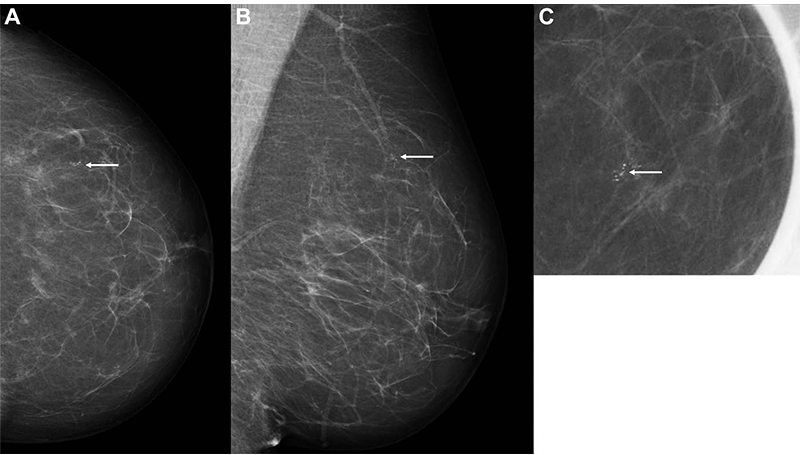
Images of a 60-year-old woman with an invasive screen-detected cancer with artificial intelligence (AI) score of 1 on the screening mammograms. (A) Mammogram of the left breast from craniocaudal view. (B) Mammogram of the left breast from a mediolateral oblique view. (C) Craniocaudal cone view mammogram with magnification. AI score is defined as the overall examination-level score from the AI system, and a score of 1 is indicative of a low probability of breast cancer and 10 of a high probability. The arrows indicate the malignancy. Image Credit: Larsen et al, Radiology 2022; 000:1-9, ©RSNA 2022
Mammograms obtained by population-based breast cancer screening programs put radiologists under a lot of stress. The use of artificial intelligence as an automated second reader for mammography has been proposed as a way to lessen this workload. Although the technology has demonstrated promising results in cancer detection, there is little evidence of its applicability in real-world screening situations.
Norwegian researchers led by Solveig Hofvind, Ph.D., from the Section for Breast Cancer Screening, Cancer Registry of Norway in Oslo, measured the effectiveness of a commercially available AI system for routine individual double reading as conducted in a population-based screening program in the new research, which is the highest of its kind to date.
BreastScreen Norway, the country’s population-based screening program, collected data from about 123,000 tests conducted on more than 47,000 women at four facilities.
There were 752 malignancies identified at screening and 205 interval cancers (cancers discovered between screening rounds) in the dataset. On a scale of one to ten, the AI system projected cancer risk, with one being the lowest risk and ten representing the highest risk. The maximum AI score of 10 was seen in 87.6% (653 of 752) of screen-detected tumors and 44.9% (92 of 205) of interval cancers.
To evaluate the AI system’s accuracy as a decision-making mechanism, the researchers devised three thresholds. The fraction of screen-detected tumors not picked by the AI system was less than 20% when using a threshold that mirrored the average individual radiologist’s percentage of positive interpretation. While the AI system performed admirably, the study’s reliance on retroactive data necessitates further investigation.
Greatest Potential May Be in Reduced Reading Volume
In our study, we assumed that all cancer cases selected by the AI system were detected. This might not be true in a real screening setting. However, given that assumption, AI will probably be of great value in the interpretation of screening mammograms in the future.
Dr. Solveig Hofvind, PhD, Section for Breast Cancer Screening, Cancer Registry of Norway
The findings revealed that screening-detected tumors with low versus high AI scores had superior histopathologic features and a better prognosis. For interval tumors, the outcomes were diametrically opposed. This could mean that interval tumors with poor AI scores are actual interval cancers that are not apparent on mammograms.
The high proportion of real negative investigations with a low AI score has the ability to decrease the interpretative volume while letting only a tiny percentage of tumors go undiagnosed. The radiologist could still detect these malignancies by utilizing AI as one of two readers in a double reading situation, according to the study.
Based on our results, we expect AI to be of great value in the interpretation of screening mammograms in the future. We expect the greatest potential to be in reducing the reading volume by selecting negative examinations.
Dr. Solveig Hofvind, PhD, Section for Breast Cancer Screening, Cancer Registry of Norway
Although additional research is needed before AI can be used in clinical breast cancer screening, the findings of the study provide a foundation for future research, especially prospective studies, according to Dr. Hofvind.
“We are looking forward to testing out different scenarios for AI using retrospective data and then running a prospective trial,” she concluded.
Journal Reference:
Larsen, M., et al. (2022) Artificial Intelligence Evaluation of 122 969 Mammography Examinations from a Population-based Screening Program. Radiology. doi.org/10.1148/radiol.212381.