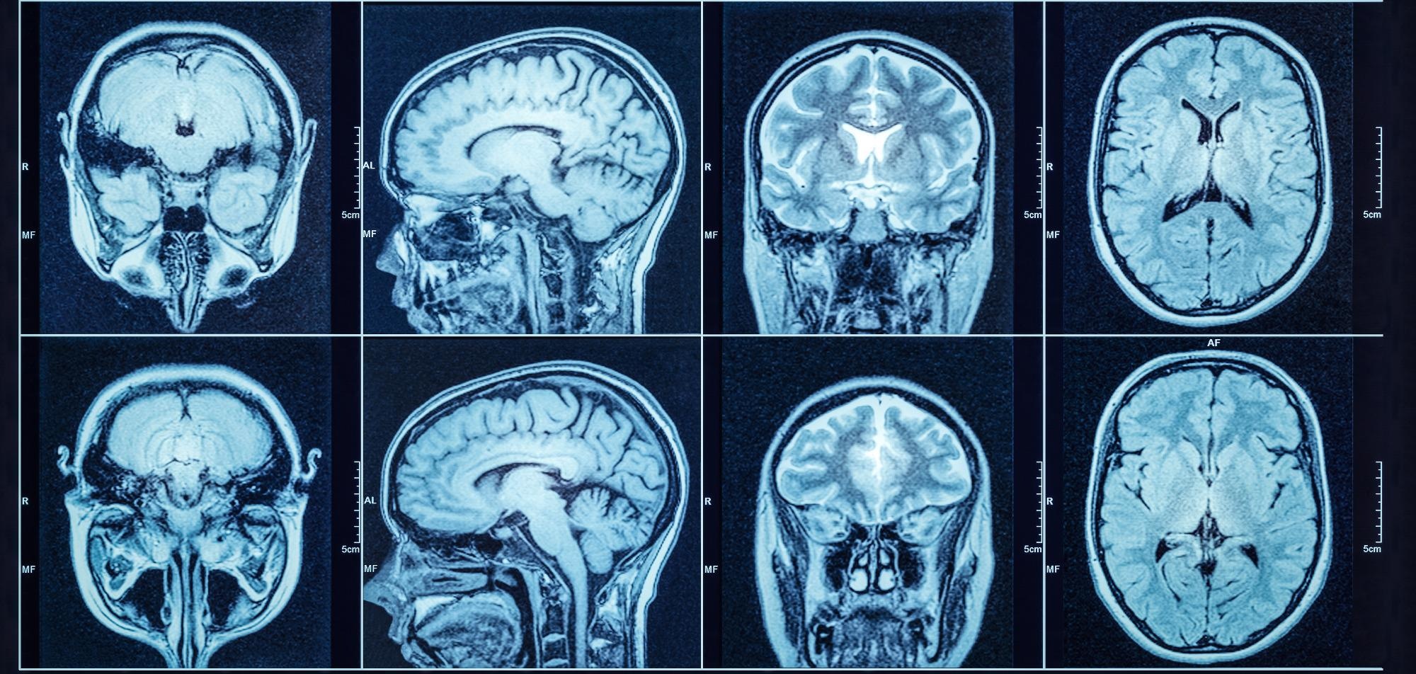
Good brain health is often a good indicator of chronological age. Today’s convolutional neural networks (CNN) are able to accurately predict a healthy patient’s age from structural MRI brain scans.

Image Credit: Shutterstock.com/Triff
A team of researchers based at the School of Biomedical Engineering & Imaging Sciences, King’s College London, has created a novel machine learning tool that examines brain MRIs and can predict brain age.
In effect, the team has designed a screening tool that is able to automatically identify brains that appear older than the chronological age in real-time, using standard clinical MRI scans.
The research published in Neuroimage demonstrates that brains suffer volume loss during the natural aging process. Given that the volume loss is correct for the patient’s age, the new machine learning tool can accurately determine the patient’s age.
Detecting Diseased Brains
When a patient’s MRI displays a brain showing signs of disease – such as dementia – there is typically a disproportionate loss of volume, and the majority of existing brain-age models have been optimized for scans that are not part of a patient’s routine examination or are dependent on costly pre-processing phases which limit in situ usage.
Thus, the King’s-based team set out to develop a framework that demonstrated the feasibility to be used as a screening tool in standard hospital examinations to identify older-looking brains in real-time.
This in turn could help improve the patient pathway by expediting care (including intervention where available), and possibly accelerate the development of disease-modifying drugs through improved clinical trial enrolment.
Dr. Thomas Booth, Senior Lecturer in Neuroimaging at King’s College London
The team believes that their tool can close the gap from the scan to review by an expert panel if available through automating such processes.
Employing the machine learning-based neuroradiology report classifier, the team was able to produce a vast dataset containing 23,302 ‘radiologically for normal age’ MRI results from two of London’s largest hospitals, Guy’s and St. Thomas’ NHS Foundation Trust and King’s College Hospital, using the pre-existing neuroradiology reports.
The team then took a novel approach. The scans were applied to an additional deep learning image algorithm with little computational pre-processing to the vast dataset they had acquired. Typical scan types obtained from a third institution were then introduced alongside an open-source dataset.
Testing the Model
Booth and his team then set about testing their model on scans that displayed disproportionate volume loss in the brain and evaluated the brain-age framework they had constructed through a series of heatmaps that highlighted areas of brain volume loss.
By using such a large dataset, the team was able to suitably train the machine learning model due to the model’s ability to generalize out-of-sample data.
Currently abnormal older-appearing brains are detected sometime after the scan at the time of reporting. The most accurate reports will be in centres where there are neuroradiologists however few centres have neuroradiologists.
Dr. Thomas Booth, Senior Lecturer in Neuroimaging at King’s College London
This indicates that the model could have significant effects on patient care and drug development, especially in smaller neurology centers where there is a lack of neuroradiology experts.
The automatic detection of volume loss in real-time could pave the way for the screening of common problems associated with neurodegeneration during scans obtained for a variety of reasons.
Subsequently, diagnosis of, for example, early-stage Dementia or Alzheimer’s disease has the potential to enhance patient care by introducing early social and medical interventions. Patients could also be recruited into drug trials at a much earlier stage.
This framework could be used to take advantage of vast hospital databases to offer invaluable new resources for the testing, clinical validation and training of medical image analysis tools beyond brain-age-based applications.
References and Further Reading
Wood, D., Kafiabadi, S., et al., (2022) Accurate brain‐age models for routine clinical MRI examinations. NeuroImage, [online] 249, p.118871. Available at: https://www.sciencedirect.com/science/article/pii/S1053811922000015?via%3Dihub
Kcl.ac.uk. (2022) New screening tool developed to automatically identify older appearing brains typical of dementia. [online] Available at: https://www.kcl.ac.uk/news/new-screening-tool-developed-to-automatically-identify-older-appearing-brains-typical-of-dementia
Disclaimer: The views expressed here are those of the author expressed in their private capacity and do not necessarily represent the views of AZoM.com Limited T/A AZoNetwork the owner and operator of this website. This disclaimer forms part of the Terms and conditions of use of this website.