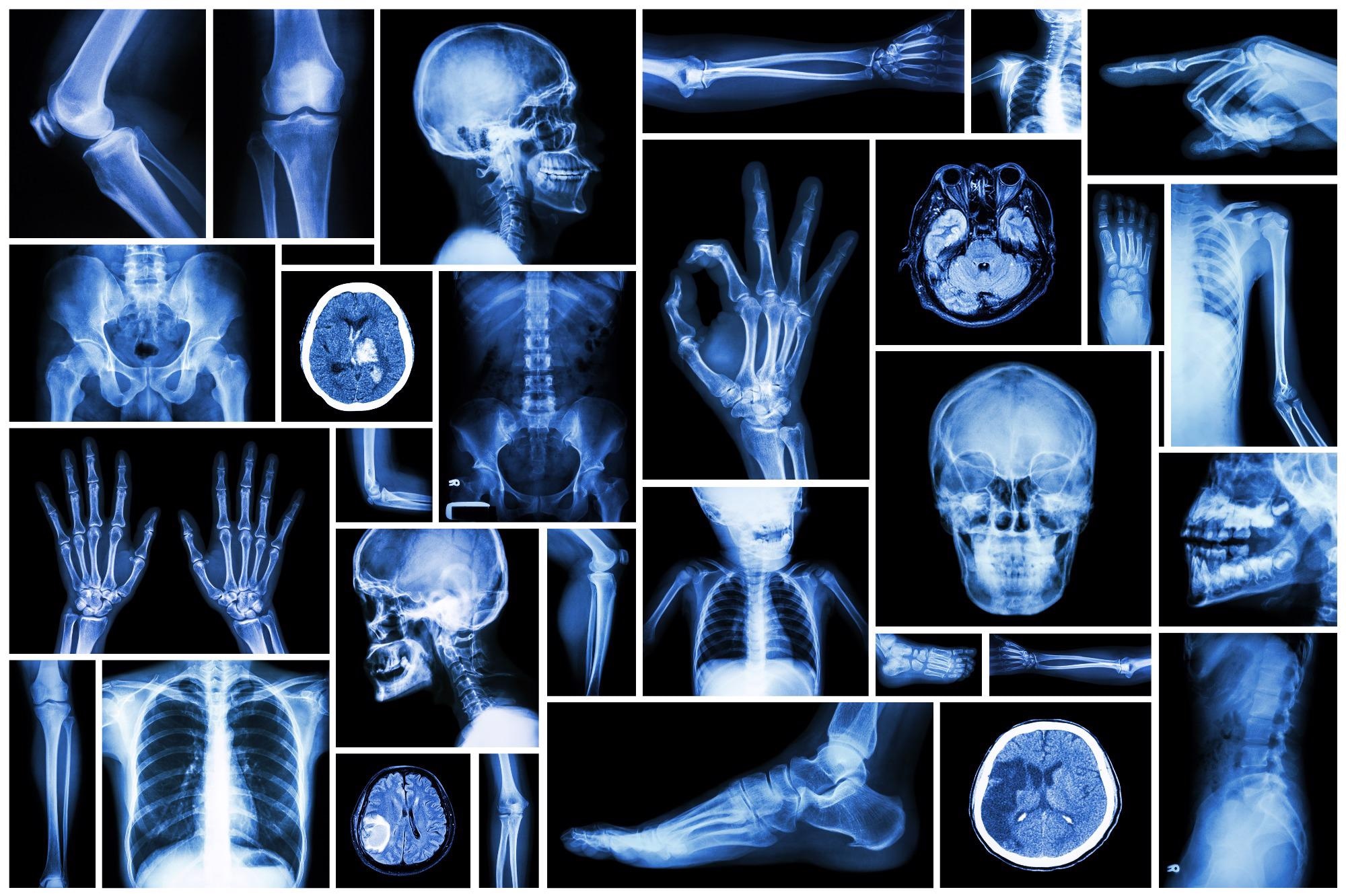
Image Credit: Shutterstock.com/ Puwadol Jaturawutthichai
Therefore, waiting for X-Rays to be assessed by radiologists can add up to this long wait time since radiologists usually interpret X-Rays for a significant number of patients. A new study has discovered that artificial intelligence (AI) can aid physicians in assessing X-Rays following an injury and suspected fracture.
Guermazi is also the Chief of Radiology, VA Boston Healthcare System.
As far as the emergency department is concerned, fracture interpretation errors constitute up to 24% of harmful diagnostic errors observed. Moreover, inconsistencies present in the radiographic diagnosis of fractures are highly common during the evening and overnight hours (5 p.m. to 3 a.m.). It is probably associated with fatigue and non-expert reading.
Using a great number of X-Rays obtained from multiple institutions, the AI algorithm called AI BoneView was trained to detect fractures of the pelvis, limbs, torso, rib cage and lumbar spine.
Expert human readers like musculoskeletal radiologists — who are subspecialized radiology doctors after obtaining focused training on reading bone X-Rays — specified the gold standard in this study and made a comparison of the performance of human readers with and without the help of AI.
A range of readers were utilized to simulate real-life scenarios, such as surgeons, radiologists, rheumatologists, orthopedic emergency physicians and physician assistants and family physicians, all of whom interpreted X-Rays in real clinical practice to diagnose fractures in their patients. Every reader’s diagnostic accuracy of fractures, with and without the help of AI, was compared to the gold standard.
Furthermore, they evaluated the diagnostic performance of AI alone in comparison to the gold standard. AI support helped increase readers’ sensitivity by 16% and decreased missed fractures by 29% and by 30% for evaluations with more than one fracture while enhancing specificity by 5%.
Guermazi hopes that AI can be a robust tool to assist radiologists and other physicians to increase efficiency and enhance diagnostic performance, while possibly enhancing patient experience at the time of clinic visit or hospital.
Our study was focused on fracture diagnosis, but similar concept can be applied to other diseases and disorders. Our ongoing research interest is to how best to utilize AI to help human healthcare providers to improve patient care, rather than making AI replace human healthcare providers. Our study showed one such example.
Ali Guermazi, MD, PhD, Study Corresponding Author and Professor of Radiology & Medicine, Boston University School of Medicine
The findings of the study have been published online in the journal Radiology.
The co-authors of the study are Chadi Tannoury, Andrew Kompel, Akira Murakami, Alexis Ducarouge, Andre Gillibert, Xinning Li, Antoine Tournier, Youmna Lahoud, Mohamed Jarraya, Elise Lacave, Hamza Rahimi, Alois Pourchot, Robert Parisien, Alexander Merritt, Douglas Comeau, Nor-Eddine Regnard, and Daichi Hayashi.
This study was financially supported by GLEAMER Inc.
Journal Reference:
Guermazi, A., et al. (2021) Improving Radiographic Fracture Recognition Performance and Efficiency Using Artificial Intelligence. Radiology. doi.org/10.1148/radiol.210937.