Mar 17 2021
Both cancer and healthy cells can appear almost the same under a microscope. One way to distinguish these cells is to examine the pH level, or the level of acidity, within them.
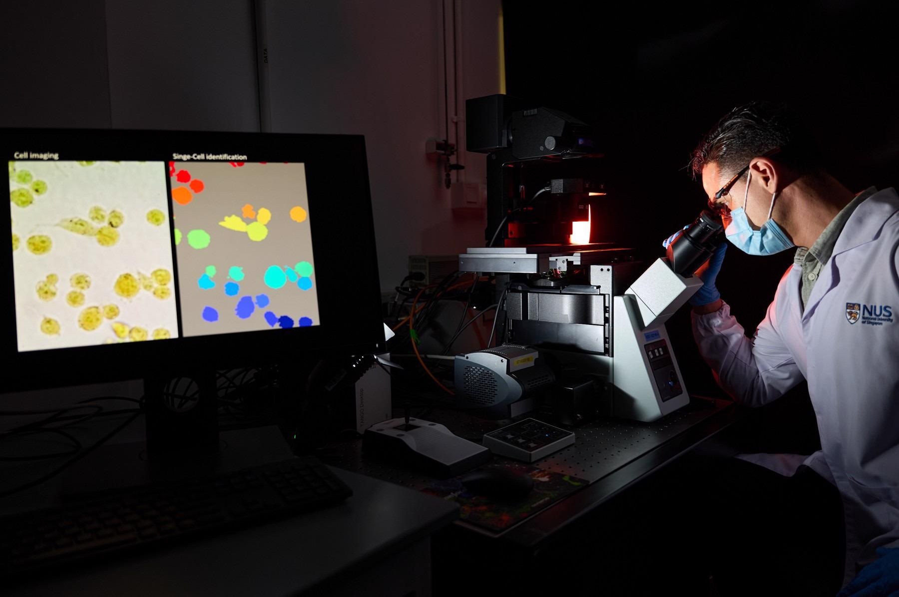
 The simple and low-cost technique developed by NUS researchers can accurately monitor and analyze living cells without causing any toxicity to the cells or the need to kill them. Image Credit: National University of Singapore.
Exploiting this unique feature, a team of researchers from the National University of Singapore (NUS) has devised a method that makes use of artificial intelligence (AI) to find out whether a single cell is cancerous or healthy by simply examining its pH level.
Every cancer test can be finished in less than 35 minutes, and single cells can be categorized with a precision speed of over 95%.
The study, headed by Professor Lim Chwee Teck, Director of the Institute for Health Innovation & Technology (iHealthtech) from NUS, was initially published in the APL Bioengineering journal on March 16th, 2021.
The ability to analyse single cells is one of the holy grails of health innovation for precision medicine or personalised therapy. Our proof-of-concept study demonstrates the potential of our technique to be used as a fast, inexpensive, and accurate tool for cancer diagnosis.
Lim Chwee Teck, Professor, Department of Biomedical Engineering, National University of Singapore
Using AI for Cancer Detection
Existing methods used for studying a single cell can cause toxic effects or may even destroy the cell. On the other hand, the new technique developed by Professor Lim and his research team can differentiate between cells that originate from cancerous and normal tissues and also among different forms of cancer.
Most significantly, all these aspects can be realized without destroying the cells.
The new method developed by the NUS team depends on using bromothymol blue—a pH-sensitive dye that shifts color based on the acidity level of a solution—onto living cells.
Because of its intracellular function, each cell type exhibits its own 'fingerprint’ containing its own special combination of red, green and blue (RGB) components when irradiated. When compared to healthy cells, tumor cells have an altered pH and, hence, tend to react differently to the dye, and this feature alters their RGB fingerprint.
With the help of a standard microscope fitted with a digital color camera, the NUS researchers captured the RGB components produced from the dye within the cells.
Then, using the newly developed AI-based algorithm, they were able to quantitatively plot the special acidic fingerprints so that the types of cells analyzed can be precisely and easily detected.
It is possible to simultaneously image an unlimited number of cells emerging from different cancerous tissues. Single-cell features can also be extracted and examined. When compared to existing standard ways of cancer cell imaging that involve many hours, the new process developed by the NUS researchers can be finished within 35 minutes.
Unlike other cell analysis techniques, our approach uses simple, inexpensive equipment, and does not require lengthy preparation and sophisticated devices. Using AI, we are able to screen cells faster and accurately. Furthermore, we can monitor and analyse living cells without causing any toxicity to the cells or the need to kill them. This would allow for further downstream analysis that may require live cells.
Lim Chwee Teck, Professor, Department of Biomedical Engineering, National University of Singapore
Opening the Door for Faster Detection
Since the new method is fast, easy, and cost-effective, and delivers high throughput, the researchers are planning to produce a real-time model of this method in which tumor cells can be automatically detected and instantly isolated for additional downstream molecular analysis, like genetic sequencing, to find out any promising drug treatable mutation.
We are also exploring the possibility of performing the real-time analysis on circulating cancer cells suspended in blood. One potential application for this would be in liquid biopsy where tumour cells that escaped from a primary tumour can be isolated in a minimally-invasive fashion from bodily fluids such as blood.
Lim Chwee Teck, Professor, Department of Biomedical Engineering, National University of Singapore
Besides this, the researchers are looking forward to improving their concept to identify different phases of malignancies from the examined cells.
Journal Reference:
Belotti., Y. et al. (2021) Machine learning based approach to pH imaging and classification of single cancer cells. APL Bioengineering. doi.org/10.1063/5.0031615.