Aug 14 2020
Researchers from the Eye Clinic of the University Hospital Bonn, Stanford University, and the University of Utah have developed an artificial intelligence (AI)-based software that enables the accurate evaluation of the progression of geographic atrophy (GA).
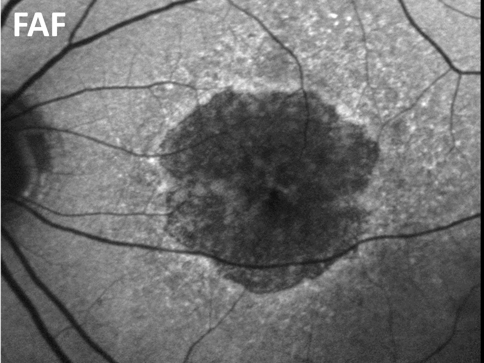
 Fundus autofluorescence (FAF): Image of the central retina in a patient with geographic atrophy—serves as a reference for optical coherence tomography (OCT). Image Credit: © University Eye Hospital Bonn.
Geographic atrophy is a disease of the light-sensitive retina that occurs due to age-related macular degeneration (AMD).
This novel technique enables fully automated measurement of the main atrophic lesions with the help of data obtained from optical coherence tomography, which offers three-dimensional visualization of the retina’s structure.
Moreover, the researchers can accurately identify the integrity of light-sensitive cells of the whole central retina, as well as detect progressive degenerative variations of what are called the photoreceptors beyond the main lesions.
The study results will be used to evaluate the effectiveness of new innovative therapeutic approaches. The study was published recently in the JAMA Ophthalmology journal.
Currently, there exists no viable treatment for geographic atrophy, which is one of the most common reasons for blindness in industrialized nations. The disease impairs the cells of the retina and destroys them.
The main lesions, regions of the degenerated retina, also called “geographic atrophy,” tend to expand with the progression of the disease and lead to blind spots in the visual field of the patient. A main difficulty faced in the case of assessment therapies is that the lesions tend to progress gradually, implying that a long follow-up period is required for intervention studies.
When evaluating therapeutic approaches, we have so far concentrated primarily on the main lesions of the disease. However, in addition to central visual field loss, patients also suffer from symptoms such as a reduced light sensitivity in the surrounding retina.
Dr Frank G. Holz, Professor and Director, Eye Clinic, University Hospital Bonn.
Dr Holz added, “Preserving the microstructure of the retina outside the main lesions would therefore already be an important achievement, which could be used to verify the effectiveness of future therapeutic approaches.”
Integrity of Light-Sensitive Cells Predicts Disease Progression
Moreover, the researchers could demonstrate that the integrity of light-sensitive cells exterior to the geographic atrophy areas is a predictor of the future progression of the disease.
It may therefore be possible to slow down the progression of the main atrophic lesions by using therapeutic approaches that protect the surrounding light sensitive cells.
Monika Fleckenstein, Professor, Moran Eye Center, University of Utah
Prof. Fleckenstein is the initiator of the Bonn-based natural history study on geographic atrophy, based on which this study was performed.
Research in ophthalmology is increasingly data-driven. The fully automated, precise analysis of the finest, microstructural changes in optical coherence tomography data using AI represents an important step towards personalized medicine for patients with age-related macular degeneration.
Dr Maximilian Pfau, Study Lead Author, Eye Clinic, University Hospital Bonn
Dr Pfau, who is currently a fellow of the German Research Foundation (DFG) and postdoctoral fellow at Stanford University in the Department of Biomedical Data Science, added that “It would also be useful to re-evaluate older treatment studies with the new methods in order to assess possible effects on photoreceptor integrity.”
Journal Reference:
Pfau, M., et al. (2020) Progression of Photoreceptor Degeneration in Geographic Atrophy Secondary to Age-related Macular Degeneration. JAMA Ophthalmology. doi.org/10.1001/jamaophthalmol.2020.2914.