Image Credit: Micha Weber/Shutterstock.com
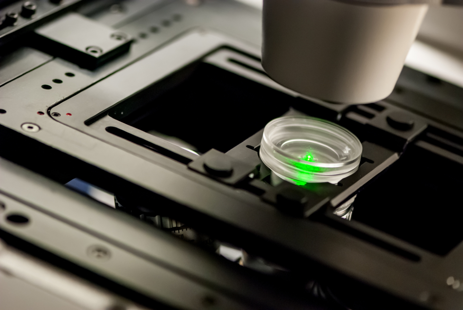
The high-resolution laser scanning confocal microscope (LSCM) is also popularly known as a laser scanning microscope. This is a highly powerful instrument for obtaining high-resolution images and three-dimensional reconstructions.
The main feature of LSCM is its ability to capture sharp images of thick specimens at various depths. Images are taken point-by-point and are then reconstructed with the help of a computer. After the images are reconstructed, various software is used to obtain high-resolution surface images with accurate nanometer measurements.
Robotic techniques have advanced in leaps and bounds. Robots are effectively utilized to observe and study microscopic samples accurately by imaging it from multiple directions. To make this possible and feasible, a device with a high degree of rotational ability has been developed. This system has been integrated with a microscope.
To image the object, the first step is to make sure that the robot's rotational axis and the sample are in line with each other. Once this crucial step is completed, multidirectional images are captured.
Advantages of Using Laser Scanning Microscopy in Robotics
- Imaging can be performed in all directions – An expert robotic arm with five degrees of freedom at the objective tip enables the microscope to access any point of a three-dimensional sample.
- Multiple areas of imaging - The miniature objective supports numerous probes to be tightly packed around a three-dimensional sample for instantaneous imaging of multiple areas of the sample.
- High resolution at a cellular level - This device can record hundreds of cells per imaged area that may be distributed over a larger surface area (example: brain cells imaging).
Applications of Laser Scanning Microscopy in Robotics
Neuroscience research
A robotic optical microscopy system has been developed by the researchers of Prof. Mark Schnitzer's laboratory.
This technology can simultaneously view and record individual areas of a single three-dimensional sample. Miniature objectives are mounted on robotic arms to move around a sample with five degrees of freedom, allowing cellular-level imaging in many places to visualize long-range interactions of cells scattered around the sample.
The technology is used in the functional studies of neuron populations throughout the brain and could be used for other basic biomedical research or surgical robotics.
Robotic-assisted endomicroscopy
Recent research has led to the development of a novel robotic device that is capable of endomicroscopy imaging.
The device contains a fast and precise scanning technology. One of the other main advantageous features of the device is its in-built image-based motion control, which enables the production of histology-like endomicroscopy mosaics.
The generated mosaics are highly magnified, which aid in conducting both in vivo and in situ cellular imaging for the study of tissue pathology. For example, the device has been used for breast-conserving surgery in humans.
As mentioned earlier, this device is robot-assisted and can surgically remove human tissues without any additional tools. The newly developed high-speed device scans 3 mm2 tissue in under 10 seconds, reducing tissue assessment time substantially.
Weld recognition
Laser scanning systems have been implemented in industrial robotic arms for accurate non-contact measurements. The laser scanner fixed on the robot runs through the shape of the object during the inspection. The application of a robot-assisted high-resolution laser scanning confocal microscope improves the precision and speed of the weld recognition.
Click here for more information on welding robotics
Soft tissue microsurgeries
Versatile laser scanning microscopy devices have been extensively used in the microsurgery of soft tissues. They are used in an array of settings that include three-dimensional printing, diagnostics, marking, and cutting.
Robot-assisted phonomicrosurgery
An improved image-based system enhanced with the ability to scan using a laser has been developed for robot-based phonomicrosurgery.
An improvement in the surgical quality of phonomicrosurgery was brought about by open-loop laser control. This system is designed to eliminate open-loop errors caused by defects in the calibration of the device or the unknown morphological structure of the sample under consideration.
This system is very accurate and reliable. It even generates a precise reading when smoke obstructs the lens of the camera. A novel image-based laser microsurgical control system was demonstrated to be highly effective with significant beneficial impacts. More precisely, the device enables microsurgeries to be safe and of high quality.
References and Further Reading
"Robotic imaging system" (U.S. Patent Application, Publication No. 20150057550)
Giataganas, P., Hughes, M., Payne, C., Wisanuvej, P., Temelkuran, B. and Yang, G. (2018). Intraoperative Robotic-Assisted Large-Area High-Speed Microscopic Imaging and Intervention. IEEE Transactions on Biomedical Engineering. PP. 1-1. Available at: https://doi.org/10.1109/TBME.2018.2837058
Dagnino, G., Mattos, L. and Caldwell, D. (2014). A vision-based system for fast and accurate laser scanning in robot-assisted phonomicrosurgery. International journal of computer assisted radiology and surgery. Available at: https://doi.org/10.1007/s11548-014-1078-9
Shen, Y., Wenfeng, W., Zhang, L., Yong, L., Lu, H. and Ding, W. (2015). Multidirectional Image Sensing for Microscopy Based on a Rotatable Robot. Sensors. 15. Available at: https://dx.doi.org/10.3390%2Fs151229872
Disclaimer: The views expressed here are those of the author expressed in their private capacity and do not necessarily represent the views of AZoM.com Limited T/A AZoNetwork the owner and operator of this website. This disclaimer forms part of the Terms and conditions of use of this website.