Researchers have confirmed pronounced constitutional varus alignment in an anatomically accurate model of the Spy II Neandertal, suggesting a deep evolutionary origin for this pattern and supporting individualized alignment in modern total knee arthroplasty (TKA). Their study, published in Scientific Reports, applied robotic-assisted TKA (i.e., knee replacement) methods to the model to explore Neanderthal biomechanics, replicating soft-tissue laxity from a comparable modern patient.
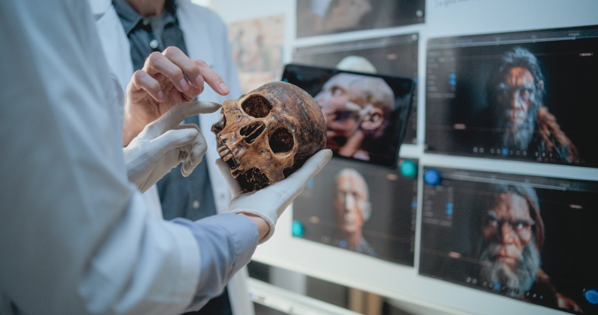 Image: Robotic-assisted knee arthroplasty methods provide insight into Homo neanderthalensis gait and the evolutionary origins of modern human knee variation. Image Credit: Frame Stock Footage/Shutterstock.com
Neandertal Knee Anatomy Challenges Modern Alignment Assumptions
Lower-limb alignment determines joint loading and is linked to degenerative joint disease, yet its evolution remains largely unexplored because no standardized framework has been applied to extinct hominins.
Fossil postcrania are mostly damaged and require reconstruction, introducing bias, and the accuracy of reconstructed alignment has never been tested. Neandertal lower-limb morphology has long provoked debate, ranging from Virchow's pathological reading to bent-hip, bent-knee gait hypotheses that were later refuted, while features such as large joint surfaces and short distal segments remain difficult to interpret.
The Spy II Neandertal is exceptionally complete, with an associated femur and tibia. This study applies robotic-assisted arthroplasty technology and the coronal plane alignment of the knee (CPAK) classification to an anatomically accurate Spy II model to characterize its alignment and explore locomotor and evolutionary implications.
Robotic Knee Surgery Technology Reconstructs Ancient Joint Mechanics
The researchers digitized casts of the Spy II right femur and left tibia using a structured-light three-dimensional (3D) scanner, generating high-resolution meshes at roughly 0.3 mm resolution.
Because only one side of each bone survives, the left tibia was mirrored to reconstruct the contralateral element, assuming bilateral symmetry of coronal alignment as seen in modern humans.
The bones were then 3D-printed at original size and checked for dimensional fidelity against the digital meshes. To simulate soft tissues, adjustable elastic elements were fixed between femoral and tibial anchor points, allowing controlled modulation of ligamentous restraint.
Laxity was calibrated to reproduce the coronal stress-strain characteristics of an adult male modern patient with matching bone alignment, sex, and skeletal maturity. The limb was mounted with the femoral head articulated against a printed acetabular model to locate the mechanical hip center, and a robotic-assisted TKA system was used in accordance with its standard workflow.
Optical trackers were pinned to the bones, landmarks were registered, and the hip, knee, and ankle centers were identified to establish the mechanical axis. Articular surfaces were mapped, and coronal balance was assessed by applying controlled varus and valgus stresses in extension and at 90° flexion. Implant size and position were planned, and mechanical, kinematic, and functional alignment strategies were simulated.
Download a free copy of this page here!
Spy II Neandertal Reveals Pronounced 9° Varus Alignment
The authors applied robotic-assisted arthroplasty planning to Spy II to characterize its alignment, joint morphology, and inferred soft-tissue laxity. The specimen showed pronounced constitutional varus with apex-distal joint-line obliquity, a phenotype considered atypical and potentially pre-arthritic in modern clinical contexts, yet present here without evidence of degenerative joint disease.
This raises the possibility that such morphology may not carry the same pathological implications given the Neandertal body plan. Prior work reported a bicondylar angle suggesting varus, but that measure follows the femoral shaft rather than the mechanical axis and cannot capture proximal femoral differences known to distinguish Neandertals from modern humans. This study instead confirms mechanical varus within a reproducible framework.
The varus exceeds typical modern values and is driven mainly by a marked tibial component. Similar phenotypes in contemporary populations are associated with intensive weight-bearing activity during growth.
The authors caution against direct behavioral analogies but note their findings are not inconsistent with evidence favoring power-based locomotion over endurance running, and with encounter-and-ambush hunting.
They frame the morphology as consistent with skeletal adaptation to mechanical loading, and interpret near-neutral trochlear orientation alongside distinctive patellar morphology as a patellofemoral joint adapted to varus-dominated loading. They also argue that Neandertal pelvic breadth should not be assumed to imply substantially greater mechanical hip width.
From an arthroplasty perspective, restoring a neutral axis resulted in substantial medial imbalance, whereas strategies that partially respect native alignment produced more balanced profiles, reinforcing the notion that constitutional alignment patterns are stable phenotypes rather than pathological deviations. Contemporary implants also mismatched the specimen's femoral proportions.
Limitations include inferred soft tissue, a single specimen, and mirrored reconstruction.
Ancient Knee Alignment Offers Clues for Modern Arthroplasty
By applying robotic-assisted total knee arthroplasty planning to an anatomically accurate Spy II model, this study demonstrates the potential of integrating robotic surgical technology with high-resolution fossil reconstructions to investigate lower-limb alignment in extinct hominins.
The specimen showed a pronounced constitutional varus alignment driven mainly by the tibial component, a phenotype considered potentially pre-arthritic in modern clinical contexts yet present here without evidence of joint disease.
The authors interpret this as suggesting a deep evolutionary origin for such alignment patterns, while cautioning that functional inferences remain speculative. The findings reinforce the view that constitutional alignment patterns are stable biomechanical phenotypes rather than pathological deviations, supporting individualized alignment in contemporary arthroplasty.
Because the analysis rests on a single specimen, with soft-tissue behavior inferred and mirrored reconstruction, the authors stress that generalization should be cautious and that further work on more specimens is needed.
Journal Reference
Pott, H., et al. (2026). Robotic-assisted knee arthroplasty methods provide insight into Homo neanderthalensis gait and the evolutionary origins of modern human knee variation. Scientific Reports, 16, 33917. DOI:10.1038/s41598-026-77544-z
https://www.nature.com/articles/s41598-026-77544-z
Disclaimer: The views expressed here are those of the author expressed in their private capacity and do not necessarily represent the views of AZoM.com Limited T/A AZoNetwork the owner and operator of this website. This disclaimer forms part of the Terms and conditions of use of this website.